
Fig. 1.
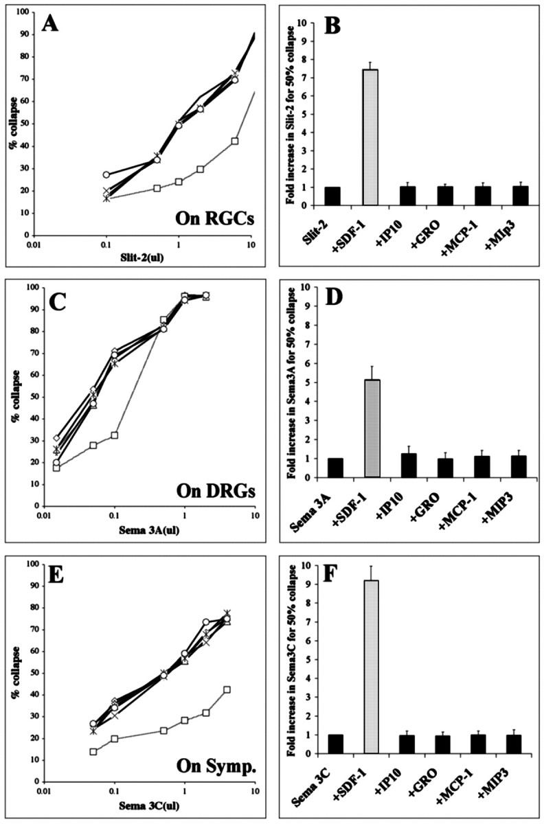
SDF-1 reduces the growth cone collapsing activities of sema-3A, sema-3C, and slit-2. A, The percentages of RGC growth cones collapsed in response to increasing concentrations of supernatant containing recombinant slit-2.Curves representing responses to slit-2 alone (open circles) or in combination with the chemokines IP-10, GROα, MCP-1, or MIP-3α are indistinguishable, indicating that slit-2 was equally potent alone or in combination with these chemokines. In the presence of SDF-1, higher concentrations of slit-2 were required to obtain a comparable collapse response (open squares, dotted line), indicating that slit-2 was less effective when combined with SDF-1. B, Fold increase in slit-2 concentration required to achieve 50% collapse in the presence of selected chemokines. This measure is equivalent to fold reduction in sensitivity to slit-2. C, The percentages of DRG growth cones collapsed in response to increasing concentrations of supernatant containing recombinant sema3A (open circles). In the presence of SDF-1, higher concentrations of sema3A were required to obtain a comparable collapse response (open squares, dotted line).D, Fold increase in sema3A concentration required to achieve 50% collapse in the presence of selected chemokines.E, The percentages of sympathetic growth cones collapsed in response to increasing concentrations of supernatant containing recombinant sema3C (open circles). In the presence of SDF-1, higher concentrations of sema3C were required to obtain a comparable collapse response (open squares,dotted line). F, Fold increase in sema3C concentration required to achieve 50% collapse in the presence of selected chemokines. All chemokines were applied at a concentration of 100 ng/ml.