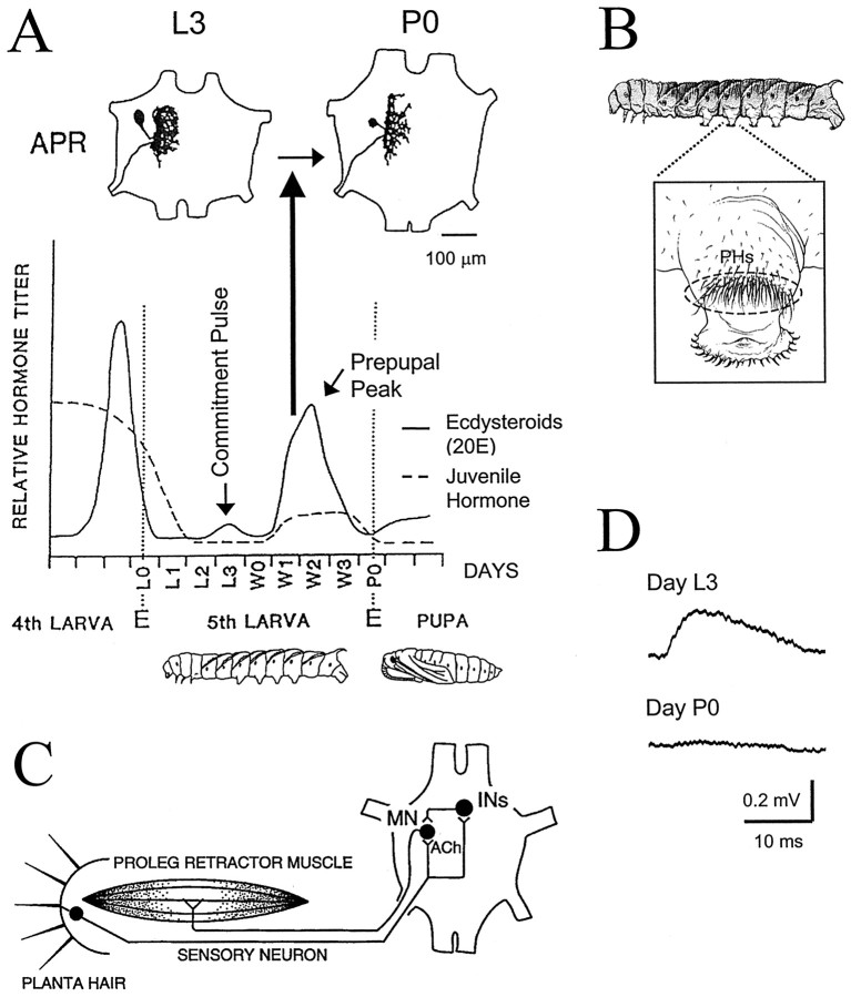
Fig. 1.
Steroid-mediated regression of APR dendrites and dismantling of the proleg withdrawal reflex during the larval–pupal transformation of Manduca. A, top, Camera lucida drawings show individual APRs stained with cobalt chloride on day L3 (left) and day P0 (right). The outline of an abdominal ganglion is shown, with the anterior end up. The APR dendritic arbor is significantly reduced on day P0 (Streichert and Weeks, 1995; Sandstrom and Weeks, 1998). The timeline illustrates changes in relative hemolymph levels of ecdysteroids (solid line) and juvenile hormone (dashed line) from the late fourth larval instar to the early pupal stage [hormone titers redrawn from Bollenbacher et al. (1981) and Riddiford and Truman (1994)]. Developmental days relevant to this study are indicated along the horizontal axis. During the fifth (final) instar, the juvenile hormone titer drops and is followed by a small rise in ecdysteroids (20E) on day L3, termed the commitment pulse, and a larger rise in 20E spanning days W1 to P0, termed the prepupal peak. The rise of the prepupal peak of 20E triggers dendritic regression in proleg motoneurons (bold vertical arrow) (Weeks and Truman, 1985, 1986; Weeks, 1987; Weeks et al., 1992). E, Ecdysis (shedding of the cuticle from the previous stage; indicated byvertical dotted lines); L0, day of ecdysis to the fifth larval instar; L1, L2, etc., days after L0; P0, day of ecdysis to the pupal stage;W0, day of wandering (when the larva ceases feeding and burrows underground); W1, W2, etc., days after wandering. B, Lateral view of a Manducalarva (anterior to the left) with the proleg in abdominal segment 4 enlarged (inset) to illustrate the dense array of PHs (enclosed by dashed oval) near the proleg tip. C, Neural circuit for the proleg withdrawal reflex in Manduca larvae. The proleg tip (left), a proleg retractor muscle (middle), and ganglion of the same abdominal segment (right; anterior is up) are shown schematically. Neurons are indicated by filled circles. Each PH is innervated by a single sensory neuron with a cell body in the proleg epidermis and an axon that projects to the ganglion. PH-SNs excite ipsilateral proleg retractor motoneurons (MN), including the APR, via monosynaptic, nicotinic cholinergic (ACh) synapses and polysynaptic pathways through interneurons (INs) (reviewed by Weeks et al., 1997). The diagram shows the reflex circuit for the left proleg in a body segment; the circuit is duplicated for the right proleg (not shown). The ability of sensory input to evoke motor output via the circuit weakens dramatically during the larval–pupal transformation (Jacobs and Weeks, 1990; Streichert and Weeks, 1995). D, The size of EPSPs produced in APRs by PH-SNs decreases during the larval–pupal transformation. Traces show intracellular recordings from an APR on day L3 (top) and a different APR on day P0 (bottom) while stimulating action potentials in a PH-SN located in the posterior region of the PH array to evoke monosynaptic EPSPs (signal-averaged from multiple trials; APR resting membrane potential set at −60 mV). No EPSP was detectable on day P0. Data are from Streichert and Weeks (1995).

