Fig. 2.

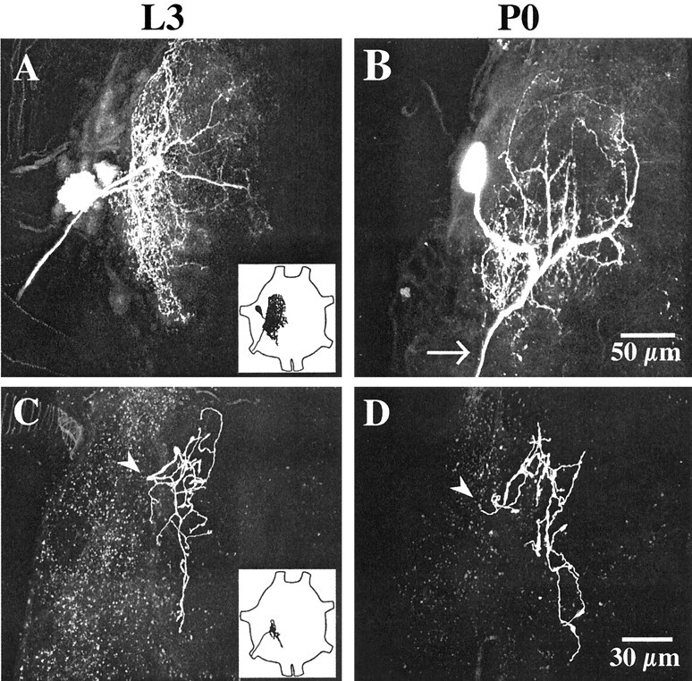
Morphology of APRs and posterior PH-SNs in larvae and pupae. Each panel is a two-dimensional projection of a stack of 0.5 μm optical sections obtained with a confocal laser scanning microscope, showing the complete extent of the central arbor of APR (A, B) or a posterior PH-SN (C, D) on day L3 (left) or day P0 (right). APRs were stained with Lucifer yellow; PH-SNs were stained with tetramethylrhodamine dextran. Anterior is up;insets show drawings of abdominal ganglia with the typical locations of an APR (A) or a posterior PH-SN (C) illustrated. A, B, The cell body of the APR appears at the left, the dendritic arbor extends to the right, toward the ganglionic midline, and the axon projects laterally and posteriorly to exit the ganglion via a segmental nerve. The variation in cell body location and neuritic branching shown here is typical (Sandstrom and Weeks, 1996). Most APR dendrites are located in dorsal and intermediate neuropil, with a less extensive projection into ventral sensory neuropil (Weeks and Jacobs, 1987). The APR dendritic arbor is less extensive on day P0 than on day L3, especially in posterior neuropil (note the loss of processes posterior to the exit of the axon from the neuropil at thearrow). C, D, The axon (arrowhead) of each posterior PH-SN enters the neuropil from a segmental nerve and arborizes within ventral sensory neuropil. The central arbors of PH-SNs map somatotopically within the CNS based on the position of the hair on the proleg (Peterson and Weeks, 1988). Posterior PH-SNs arborize in the middle and posterior regions of ipsilateral neuropil. PH-SN arbors are similar in extent on days L3 and P0. Scale bars: (in B) A, B, 50 μm; (in D), C, D, 30 μm.
