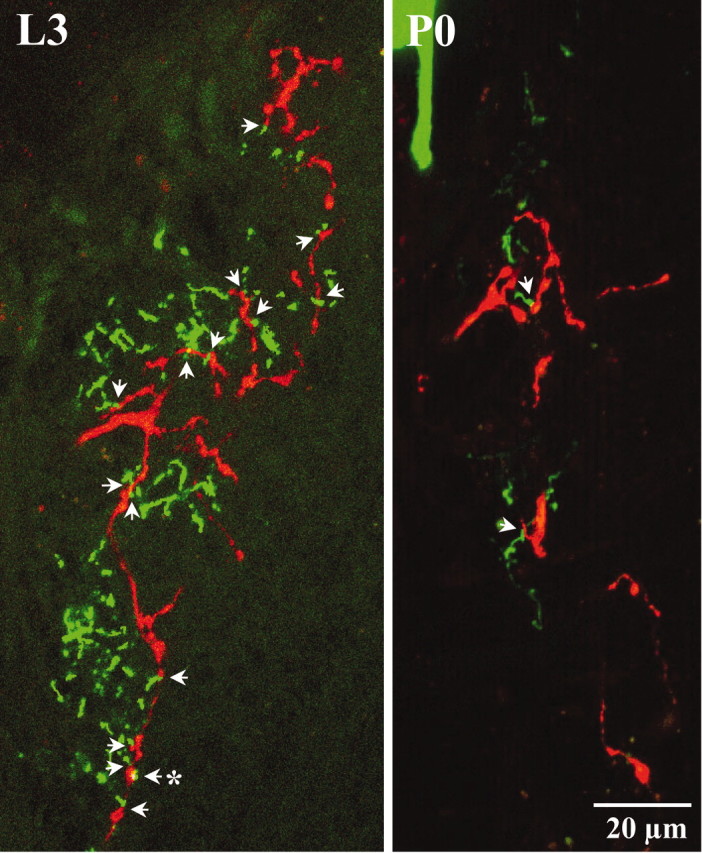
Fig. 4.

Anatomical relationship between processes of posterior PH-SNs and APRs. Each panel shows a two-dimensional projection of five sequential frames (equal to 3.4 μm depth of neuropil) containing a Lucifer yellow-stained APR (green) and a rhodamine-stained posterior PH-SN (red). Images are oriented as in Figure 2 (anteriorup). Counts of indistinguishably close anatomical juxtapositions scored as appositions in single confocal frames were converted to counts of putative synaptic contacts (arrows; see Materials and Methods). In this example, there were 15 putative synaptic contacts (represented by 21 appositions; data not shown) on day L3 and two putative synaptic contacts (represented by 4 appositions; data not shown) on day P0. Anasterisk marks the putative synaptic contact shown at higher magnification in Figure 5A. The scale bar refers to both panels.
