
Fig. 4.
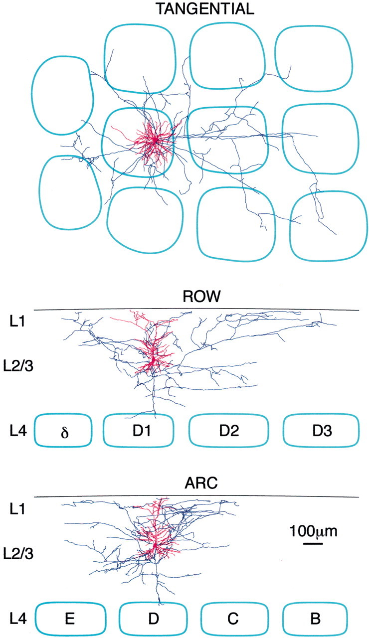
Three-dimensional reconstruction of the dendritic and axonal arbors of a layer 2/3 pyramidal neuron. During in vivo whole-cell recordings of layer 2/3 pyramidal neurons simultaneous with VSD imaging, the neuron is filled with biocytin. Subsequent staining of fixed tangential slices reveals the axonal and dendritic structure, which can be traced in three dimensions with the aid of a computer. The barrel field is visualized by cytochromec staining. The axon extends into all neighboring barrel-columns, whereas the dendrites remain close to the soma. The axon extends farther along the row than along the arc.