
Fig. 2.
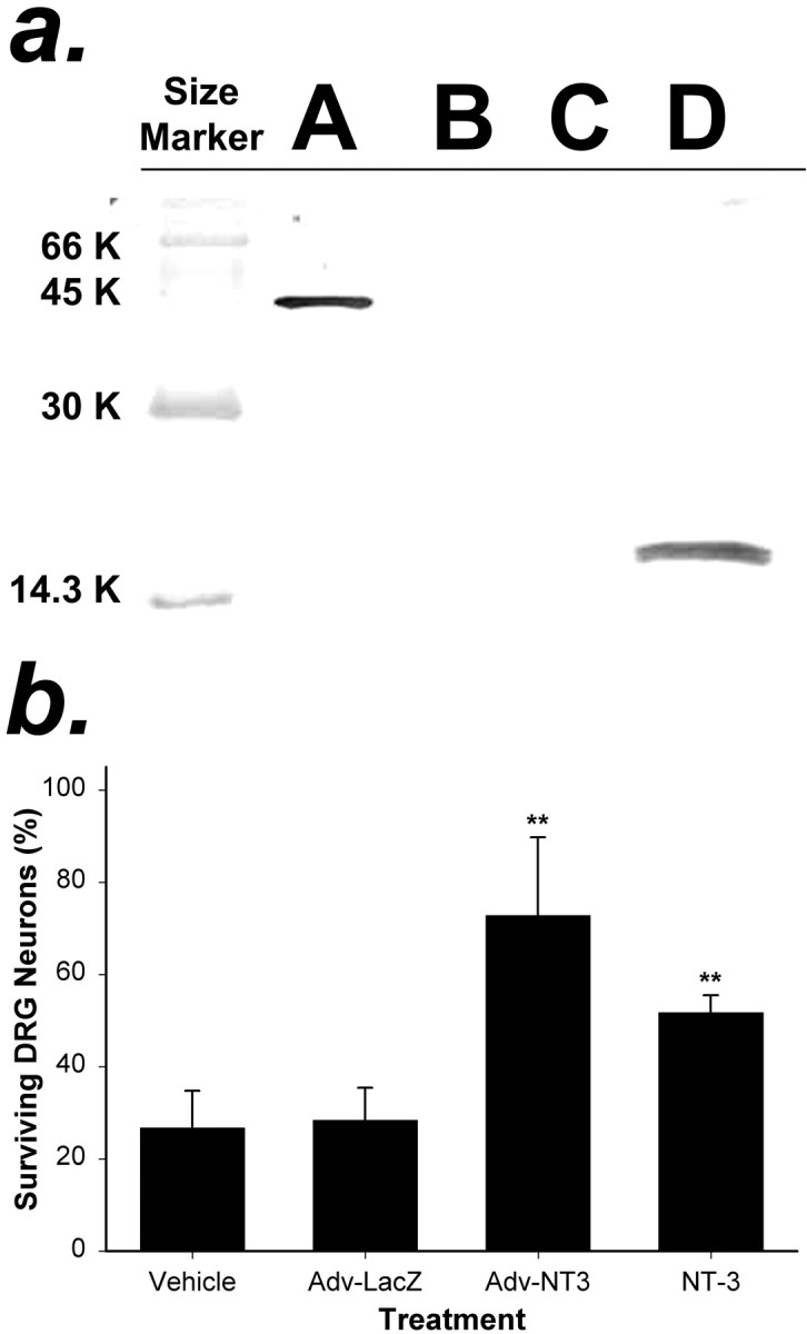
Analysis of NT-3 protein produced by cells transduced with Adv.EFα-NT3 in vitro.a, Western blot analysis of the conditioned medium of HeLa cells transduced with Adv. HeLa cells were transduced with Adv.EFα-NT3 or Adv.EFα-LacZ at an MOI of 100. After 12 hr, the medium was replaced with serum-free medium. After 48 hr, the conditioned medium was collected. Western blot analysis using an anti-FLAG antibody was performed to detect NT-3-FLAG in the medium.Lane A, BAP-FLAG; lane B, medium from untransduced HeLa cells; lane C, medium from HeLa cells transduced with Adv.EFα-LacZ; lane D, medium from HeLa cells transduced with Adv.EFα-NT3. A prominent band that cross-reacted with the anti-FLAG antibody is visible inlane D corresponding to the predicted size of the NT-3-FLAG hybrid protein. b, Conditioned medium of HeLa cells transduced with Adv.EFα-NT3 supported the survival of DRG neurons. Primary cultures of DRG neurons were cultured in test media for 48 hr, and the number of surviving neurons was counted in 10 fields. The test media were as follows: Vehicle, 25% conditioned medium from cultures of untransduced HeLa cells;Adv-LacZ, 25% conditioned medium from cultures of Adv.EFα-LacZ-transduced HeLa cells;Adv-NT3, 25% conditioned medium from cultures of Adv.EFα-NT3-transduced HeLa cells; and NT-3, 100 ng of NT-3 protein per milliliter of culture medium. The values are means ± SD of three wells; **p < 0.01 (ANOVA followed by the Student–Newman–Keuls test).