Fig. 6.

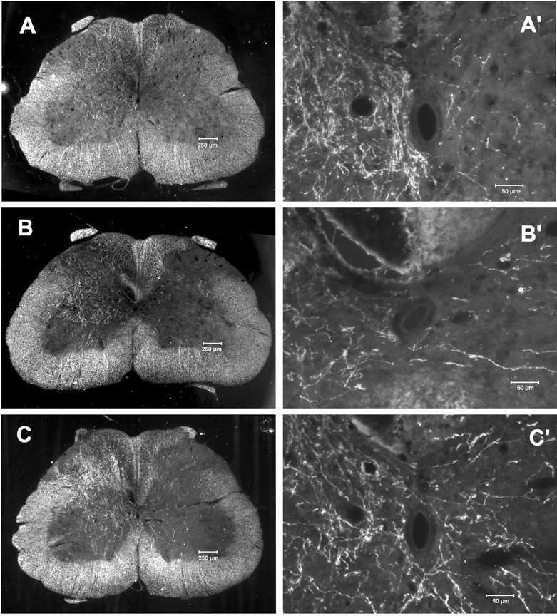
Sprouting of CST axons across the midline in the spinal cord after CSTL and Adv.EFα-NT3 transduction of motoneurons. Rats with unilateral CSTL were treated with Adv.EFα-NT3 or Adv.EFα-LacZ, whereas the unlesioned CST was labeled with BDA. Dark-field photomicrographs of spinal cord cross sections showed the unlesioned CST axons. A, Section from a normal rat (sham surgery). B, Section from an Adv.EFα-LacZ-treated rat. C, Section from an Adv.EFα-NT3-treated rat. A′–C′, Higher-power photomicrographs of the regions around the central canal.C, BDA-labeled CST neurites can be seen arising from the intact CST, traversing the midline, and growing into the gray matter of the lesioned side of the spinal cord.
