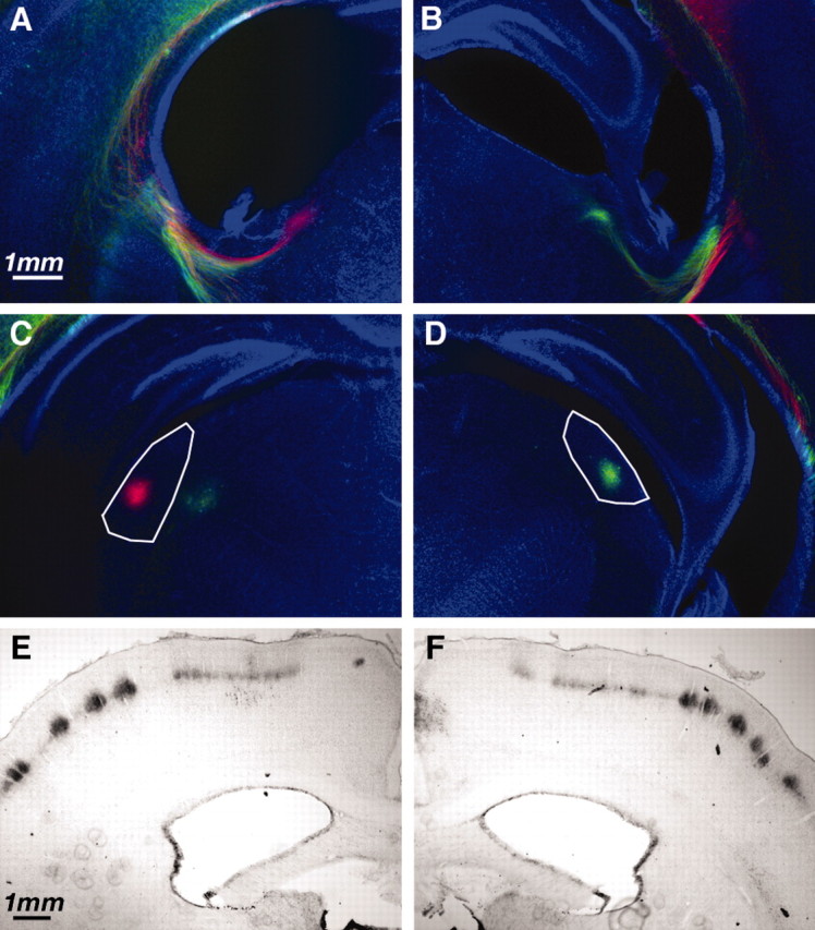
Fig. 4.

Thalamocortical projection to somatosensory and visual cortex after P2 H-I. A–D, Thalamocortical and corticothalamic axons labeled with DiI and DiD crystals placed in auditory or visual cortex. Coronal sections at the level of internal capsule (A, B) or lateral geniculate nucleus (C, D) demonstrate labeled fibers and cell bodies. To distinguish ipsilateral/H-I hemisphere from contralateral/hypoxic hemisphere, the dyes were placed in opposite configuration. For this example, the ipsilateral hemisphere (A, C) had DiI (red) placed in visual cortex and DiD (green) placed in auditory cortex. The contralateral hemisphere (B, D) had DiD (green) placed in visual cortex and DiI (red) placed in auditory cortex. Axons can be seen traversing the internal capsule (A, B), and neurons in lateral geniculate (visual thalamus) are back-labeled (C, D) in an identical manner in the two hemispheres. E, F, Cytochrome oxidase staining of sensory thalamocortical axons in patchy representation of whisker barrels in hemisphere receiving H-I (E) and hypoxia alone (F).
