
Fig. 3.
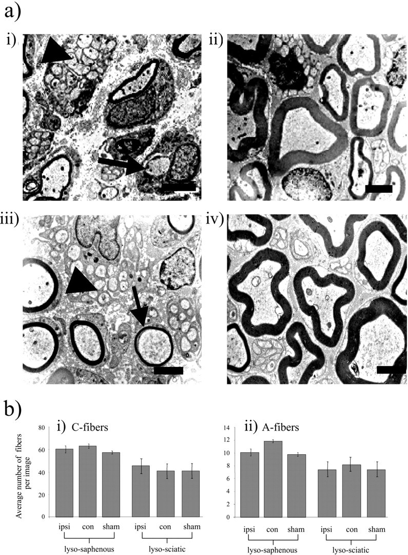
Effects of lysolecithin treatment on morphology of saphenous and sciatic nerve fibers at the EM level. a, Electron microscopy images of either the lysolecithin-treated saphenous (i) or sciatic (iii) nerves, compared with the equivalent sham-operated nerves [saphenous (ii) and sciatic (iv) nerves] on day 13 after treatment (n = 4 in each case). Thirteen days after lysolecithin treatment, ipsilateral nerves display complete demyelination in ∼40% of A-fibers with partial demyelination of many remaining myelinated fibers. The images demonstrate marked lysolecithin-induced reduction in the myelination of A-fibers (arrow), whereas C-fibers (arrowhead) remain intact (5000×). Scale bar, 2 μm. b, i andii show the mean number of C- and A-fibers recorded per image area in different conditions. Unmyelinated axons were identified as small-diameter fibers devoid of any myelin sheath (which were present in bundles, surrounded by Schwann cell cytoplasm) and were individually counted for all groups. A-fibers (with or without lysolecithin treatment) were identified as the larger-diameter axons with myelin sheaths or smaller-diameter thinly myelinated fibers or occasionally those with no myelin that were solitary. Data for both fiber types are presented as average numbers of fibers per 292 μm2 image ± SEM. A Kruskal-Wallis one-way ANOVA on ranks showed no statistically significant differences between treatment groups.