
Fig. 2.
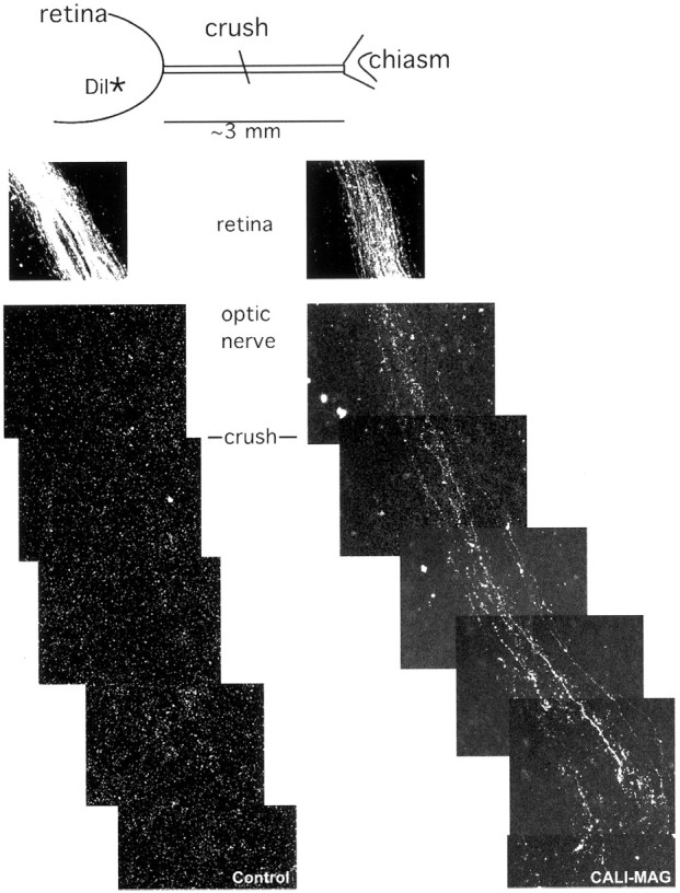
CALI of MAG enhances regeneration in E15 optic nerve. E15 retinas were explanted with attached optic nerve and tracts and given a crush injury with cold forceps halfway between the retina and chiasm. The axons were anterograde labeled with DiI crystals placed in the retina >0.5 mm from the optic fissure. As the explant was incubated for 3 d, the dye was transported up axons, and in the case of CALI-MAG (right panels), these axons could be seen beyond the point of lesion. In contrast, the control explant treated with CALI-PLP (left panels) showed no axon labeling beyond the lesion. Scale bar, 20 μm.