Fig. 5.
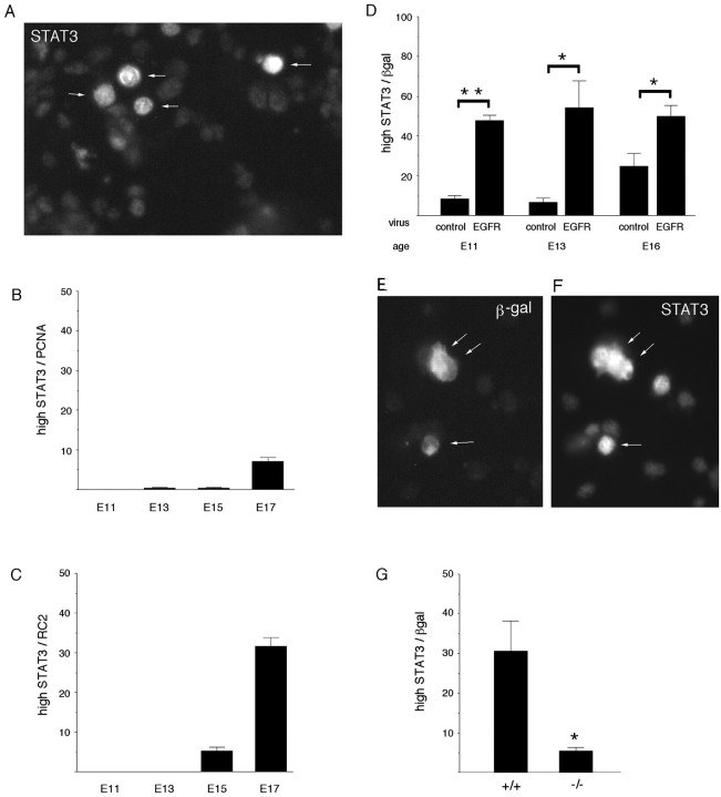
STAT3 expression changes during development and is regulated by EGFRs. A, E17 dorsolateral cortex was stained for STAT3. Note that four cells (arrows) express a high level of STAT3 immunoreactivity, whereas the majority express intermediate or low levels (see Results for quantification of the populations). The population of cells that expresses a high level of STAT3 appears at E15 and increases in size over 2 d, coexpressing the progenitor markers PCNA (B) or RC2 (C). D, Premature elevation of EGFRs induces a high level of STAT3 prematurely. Cells infected with the EGFR virus at E13 express the viral marker β-gal (E; arrows) and a high level of STAT3 (F; arrows). G, The size of the population of cells that expresses a high level of STAT3 is reduced in EGFR-null cortical explants (−/−), compared with wild type. *p ≤ 0.02; **p < 0.0001.

