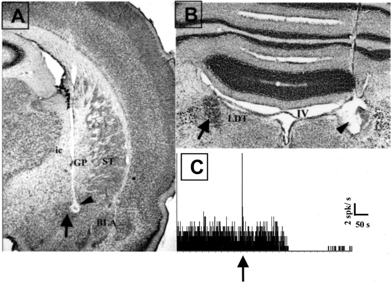
Fig. 1.
Histological and pharmacological control of electrode placement. A, Histological verification of the recording site in the CeN (arrow). The small lesion (arrowhead) in the CeN was made by passing DC current between the tips of the stimulating electrodes. Note the descending tract of the electrode through the internal capsule (ic) and the globus pallidus (GP). ST, Striatum.B, Histological verification of the recording site in LC. Note on the left the intact LC, the darkly stained nucleus just under the fourth ventricle (IV), as indicated by the arrow. On the contralateral side is the lesioned site of the LC (arrowhead) made by passing DC current between the tip of the recording electrode and the ground. The electrode tract can be seen above the lesion in the cerebellum. C, Example of a pharmacological verification of the recording from a noradrenergic neuron. Note the total inhibition of activity of the unit after systemic injection of clonidine (40 μg/kg) indicated by the arrow. All LC units and multiunit activity responded with total but transient inhibition after clonidine injection. The peak reflects the phasic activation of LC neurons during the injection.

