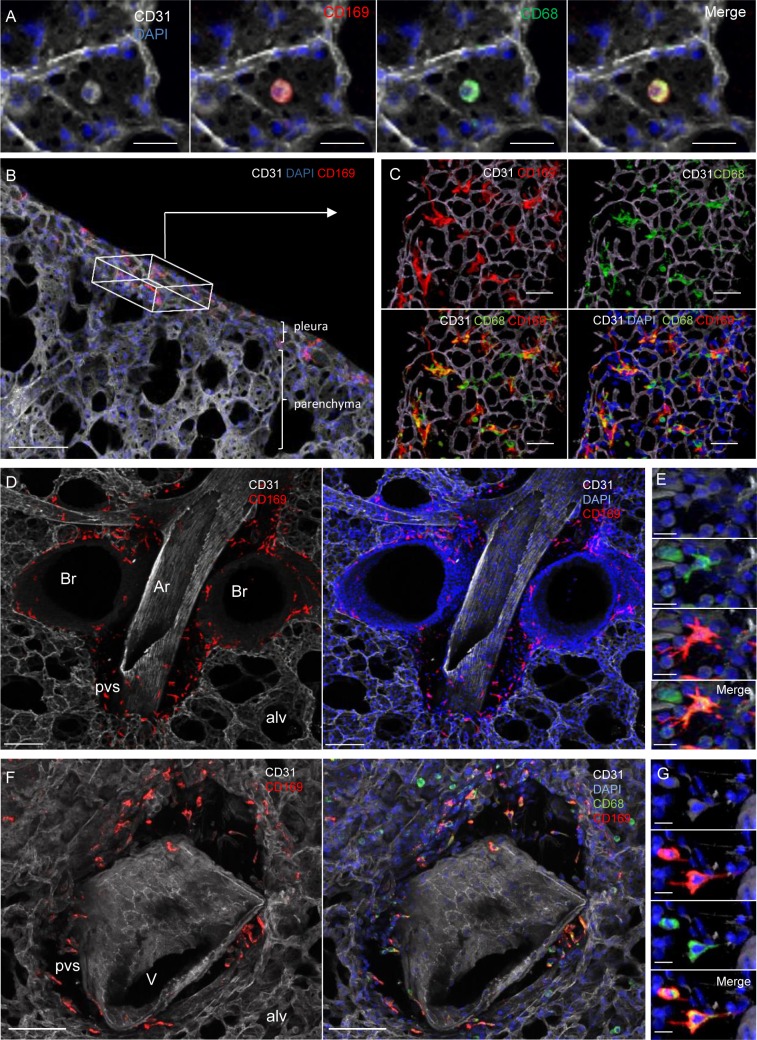
Fig 8. CD169 expression on resident macrophage populations in specific lung locations.
Lungs were recovered from naive and L. sigmodontis infected WT, ΔdblGata1 and Il-4ra-/-/Il-5-/- BALB/c mice at 70 days p.i.. Precision cut lung slices (PCLS) were prepared for confocal microscopy analysis. Representative maximum intensity projection from confocal z-stacks (z = 50μm) of PCLS stained with DAPI (blue) and fluorochrome-conjugated anti-CD31 (white), -CD68 (green) and -CD169 (red) antibodies; (A) Alveolus (surrounded by CD31+ capillaries) containing a CD68+CD169+ alveolar macrophage; Left: DAPI and CD31 channels; Middles: DAPI and CD31 with CD169 and CD68 single channels respectively; Right: Merging of the different channels showing the coexpression of CD169 and CD68 in alveolar macrophages; Scale bar = 20μm; (B) 50μm stack view of lung periphery showing CD169+ cells (red) in the visceral pleura. The box indicates pleura orientation. Scale bar = 100μm; (C) Top view of the visceral pleura showing CD169 and CD68 expression macrophages close to CD31+ capillaries. Top: CD169 and CD68 single channels with CD31. Bottom: Merging of the different channels with DAPI. Scale bar = 20μm; (D) CD31+ artery surrounded by a perivascular space (pvs) and adjacent to bronchi. Pvs and peribronchial space contain CD169+ cells. Scale bar = 100μm. (E) Higher magnification of a macrophage in a pvs around an artery showing expression of CD68 (green) and CD169 (red). Scale bar = 10μm; Top: DAPI and CD31 channels; Middle: CD169 and CD68 single channels with DAPI and CD31; Bottom: Merging of the different channels showing the coexpression of CD68 and CD169 in macrophages in artery pvs (F) CD31+ vein (identified by round nuclei and absence of adjacent bronchus) surrounded by a perivascular space (pvs) containing CD68+CD169+ cells. Scale bar = 100μm (G) Higher magnification of a macrophage in a pvs around a vein showing expression of CD68 (green) and CD169 (red). Scale bar = 10μm. Top: DAPI and CD31 channels; Middle: CD68 and CD169 single channels with DAPI and CD31; Bottom: Merging of the different channels showing the coexpression of CD169 and CD68 in macrophages in vein pvs. br: bronchus; alv: alveolus; Ar: artery; V: vein; pvs: perivascular space.

