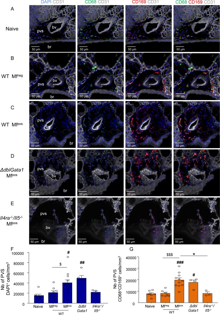
Fig 9. IL-4R/IL-5 dependent increase of lung-resident CD169+ macrophages in microfilaremic mice.
Lungs were recovered from naive and L. sigmodontis infected WT, ΔdblGata1 and Il-4ra-/-/Il-5-/- BALB/c mice at 70 days p.i.. Precision cut lung slices (PCLS) were prepared for confocal microscopy analysis. (A-E) Representative maximum intensity projection from confocal z-stacks (z = 50μm) of PCLS stained with DAPI (blue) and fluorochrome-conjugated anti-CD31 (white), -CD68 (green) and -CD169 (red) antibodies. Analysis of perivascular space (PVS) cellular content in the different groups of mice: Left column: DAPI and CD31 channels; Middle columns: DAPI and CD31 with CD169 and CD68 single channels respectively; Right column: Merging of the different channels showing CD68+CD169+ macrophages in PVS; (A) PVS of a naive uninfected mouse (B) PVS of a WT Mfneg mouse; (C) PVS of a WT Mfpos mouse; (D) PVS of a ΔdblGata1 Mfpos mouse; (E) PVS of a Il4ra-/-/Il5-/- Mfpos mouse; (F-G) Quantification of PVS cellular content. PVS cells were counted and PVS volume was measured over the 50μm z-stack (using IMARIS software) to evaluate cell concentrations. (F) total number of cells (DAPI+) per mm3 of PVS and (G) number of CD68+CD169+ macrophage per mm3 of PVS. Results are expressed as mean ± SEM (pool of 1–3 experiments) of n = 7 naive, n = 7 WT Mfneg, n = 11 Mfpos, n = 11, n = 4 ΔdblGata1 Mfpos, n = 4 Il-4ra-/-/Il-5-/- Mfpos. For each mouse, 2–3 pvs were analyzed. Kruskal-Wallis followed by a Dunns multiple comparison test: #p<0.05, ##p<0.01, ###p<0.001 represent differences between infested groups and the naïve group; $p<0.05, $ $ $p<0.001 represent differences between Mfneg and Mfpos mice *p<0.05 represent difference between Mfpos groups. bv = blood vessel; br = bronchus.

