
Fig 4. Periodic distribution of other ER markers and ER-resident proteins along peripheral ER tubules.
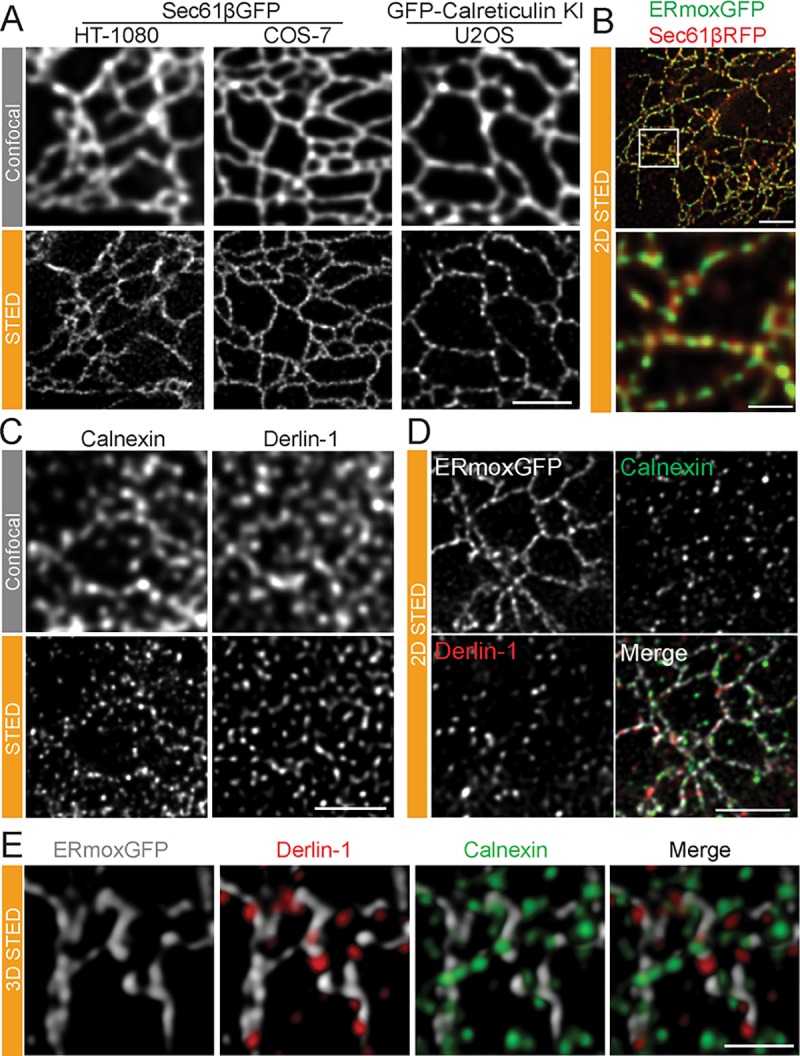
(A) Representative confocal and STED images of live HT-1080 and COS-7 cells overexpressing Sec61βGFP or live knock-in U2OS cells expressing GFP-calreticulin at endogenous levels. Scale bar, 2 μm. (B) STED images of fixed HT-1080 cells expressing lumenal ERmoxGFP (green) and membrane Sec61βmRFP (red) show distinct periodicity of these two ER reporters along peripheral ER tubules. Scale bar, 2 μm; zoom, 0.5 μm. (C) Untransfected HT-1080 cells labeled for derlin-1 or calnexin imaged by confocal and STED. Scale bar, 2 μm. (D) Association of ER-resident proteins derlin-1 and calnexin with ERmoxGFP-labeled peripheral ER tubules in HT-1080 cells by 2D STED. Scale bar, 2 μm. (E) Association of ER-resident proteins derlin-1 and calnexin with ERmoxGFP-labeled peripheral ER tubules in HT-1080 cells by 3D STED. Scale bar, 1 μm. ER, endoplasmic reticulum; ERmoxGFP, ER monomeric oxidizing environment-optimized green fluorescent protein; KI, knock-in; STED, stimulated emission depletion; 2D, two-dimensional; 3D, three-dimensional.