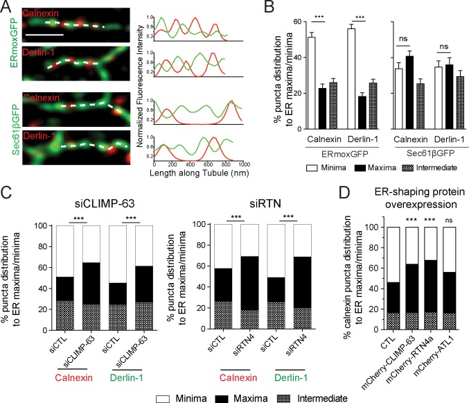
Fig 5. ER-resident proteins calnexin and derlin-1 are enriched in nanodomains depleted of lumenal ERmoxGFP.
(A) Representative merged images of single peripheral ER tubules expressing ERmoxGFP or Sec61βGFP labeled for calnexin or derlin-1. The dashed line indicates the site of line scan analysis along tubule. Fluorescence intensities of ER reporter (green) and protein (red) from line scans are presented as graphs. Scale bar, 0.5 μm. (B) Based on line scan analysis of peripheral ER tubules, percent localization of calnexin and derlin-1 puncta to ERmoxGFP or Sec61βGFP maxima and minima was quantified. Values plotted are mean ± SEM from three independent experiments (40 tubules per repeat) with one-way ANOVA for significance. ***P < 0.001. Numerical values that underlie the graphs are shown in S1 Data. (C) Based on line scan analysis of peripheral ER tubules, percent localization of calnexin and derlin-1 puncta to ERmoxGFP maxima and minima was quantified in cells transfected with siCTL, siCLIMP-63, or siRTN4. Significance was assessed by χ2 test from three independent experiments (20–40 tubules per repeat). ***P < 0.001. Numerical values that underlie the graphs are shown in S1 Data. (D) Based on line scan analysis of peripheral ER tubules, percent localization of calnexin puncta to ERmoxGFP maxima and minima was quantified in HT-1080 cells cotransfected with mCherry-CLIMP-63, mCherry-RTN4a, or mCherry-ATL1 compared with CTL. Significance assessed by χ2 test from three independent experiments (40 tubules per repeat). ***P < 0.001. Numerical values that underlie the graphs are shown in S1 Data. ATL, atlastin; CLIMP-63, cytoskeleton-linking membrane protein 63; CTL, control; ER, endoplasmic reticulum; ERmoxGFP, ER monomeric oxidizing environment-optimized green fluorescent protein; ns, not significant; RTN4a, reticulon4a; siCLIMP-63, siRNA to CLIMP-63; siCTL, siControl; siRTN4, siRNA to RTN4.

