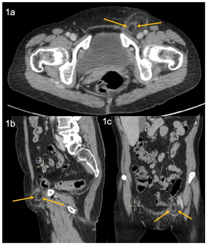
Figure 1.
65-year-old female with epiploic appendagitis within a left sided femoral hernia
FINDINGS: Fat density ovoid appendage surrounded by a high attenuation rim (arrows) and inflammatory stranding within a groin hernia with a ‘funnel shaped’ neck, protruding through the femoral ring, a) axial; b) sagittal; c) coronal. On the sagittal and coronal images the attachment of the fat density structure to sigmoid colon is evident with the thrombosed central vein seen as a high density line on the coronal view.
TECHNIQUE: Portal Venous phase IV contrast enhanced (100 ml Omnipaque) volume acquisition 128 detector CT (GE Optima CT660) of the abdomen and pelvis acquired at 1 mm slice thickness, 120 kVp 120, 50–200 mAs.

