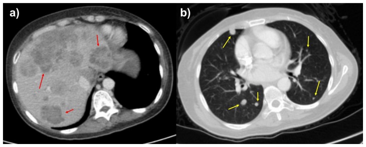
Figure 6.
48-year-old female with leiomyosarcoma of the IVC, two-year follow-up scan.
FINDINGS: Axial contrast enhanced two-year follow-up CT of the abdomen in the portal venous phase (6a) and CT of the chest in the arterial phase (6b) demonstrate worsening liver and lung metastases. The largest liver lesion measures up to 9.0 cm (initially 3.0 cm), and the largest pulmonary nodule measures up to 1.2 cm (initially 0.5 cm).
TECHNIQUE: Axial CT with sagittal and coronal reconstructions, 151 mAs (Figure 6a), 133 mAs (figure 6b), 120 kV, 3 mm slice thickness, 80 mL Omnipaque 350 intravenous contrast and 300 mL of Gastroview oral contrast.

