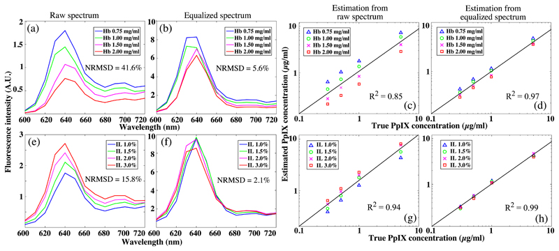
Fig. 4.
Representative spectrum of phantoms with the same PpIX concentration (0.3 μg/ml) demonstrates diverged emission intensities in (a) Hb-variable group and (e) IL-variable group, with the normalized NRMSD of 41.64% and 15.8%, respectively. The equalized spectra of both phantom groups have unified emission intensity, resulting in markedly reduced NRMSD of (b) 5.6% and (f) 2.1% (f), respectively. For both phantom set A and set B, (d, h) PpIX concentrations that were estimated from Eq. (1) show increased correlation coefficient compared with (c, g) the raw spectra.

