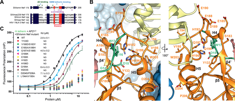
Figure 5. Structural analysis of the SIVsmm Nef tetherin binding pocket.
(A) Sequence alignment of Nef dileucine loops between SIVsmm Nef and other Nef species. Key residues in SIVsmm Nef for tetherin and AP-2 binding are highlighted. (B) Ribbon representation of SIVsmm Nef mutations investigated with SMM tetherin GDIW motif shown for reference. (C) FP assay indicates that Nef mutations in multi interfaces impair tetherin binding. See also Figure S5

