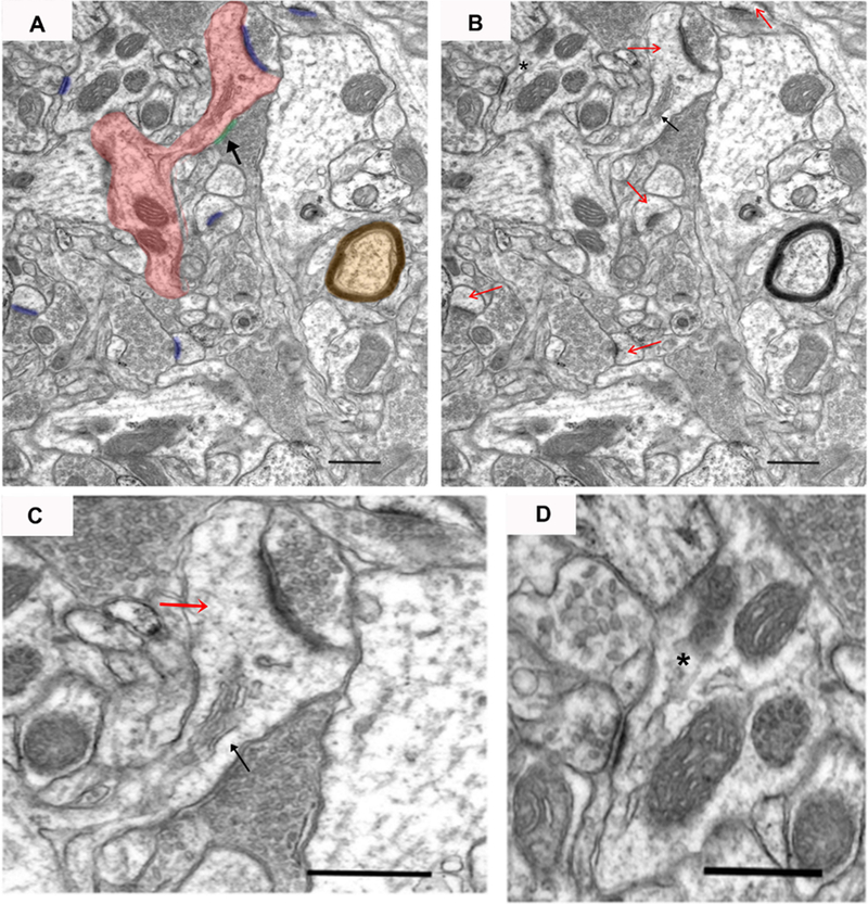
Figure 3.

An example electron micrograph of neuropil from which synapses were measured. In panel A, a dendritic spine that connects back to the dendritic shaft (dendrite) is highlighted in red, asymmetric synapses are highlighted in blue, symmetric synapse is highlighted in green with black arrow, and a myelinated axon is highlighted in orange. In panel B, red arrows mark dendritic spines, the black arrow marks a spine apparatus, and asterisk marks a postsynaptic dendrite. Panel C shows an enlarged image of a dendritic spine (red arrow) with asymmetric and symmetric synapses. Panel D shows an enlarged image of an asymmetric synapse onto a dendrite (asterisk) with a clearly defined synaptic cleft. Scale bar represents 0.5 μm.
