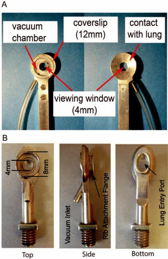
Figure 1. Evolution of The Modern Thoracic Suction Window.
A._Photographic schematic of a thoracic imaging window designed to lay directly over lung, exposed through resection of ribs. Low pressure (20 mmHg) vacuum is applied through inlet to temporarily stabilize lung tissue against the coverslip. Reprinted with permission[24]. B. Revised thoracic imaging window design incorporating an intracostal flange enabling placement directly between two ribs without need for resection. The modified window design allows for increased image stability with reduced surface exposure of the lung. Reprinted with permission[7].

