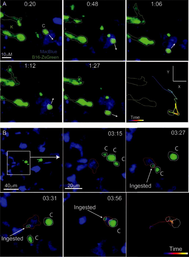
Figure 2. Live Imaging of The Early Pulmonary Metastatic Niche.
A. LIVM imaging of a ZsGreen+ B16F10 melanoma cell (Green, labeled K for karyoplast) releasing microparticles (labeled C for their alternate name cytoplasts) into the pulmonary vasculature of the myeloid reporter mouse MacBlue. In this mouse, monocytes and their progeny as well as neutrophils are labeled with CFP. White arrows show the trajectory of the myeloid cell at each timepoint as it migrates autonomously through the vasculature. The last frame of this series shows the entire path of the monocyte over the imaging time. B. LIVM of tumor microparticle (Green) being encountered and subsequently phagocytosed by a CFP+ myeloid cell (blue) in the pulmonary vasculature between 3 and 4 hours following the entry of the parental tumor cell into the lung. Microparticles are outlined in white and ingesting myeloid cell in red. The final frame of the image shows the path taken by the ingesting myeloid cell throughout the imaging time. Both A and B are reprinted with permission from[7].

