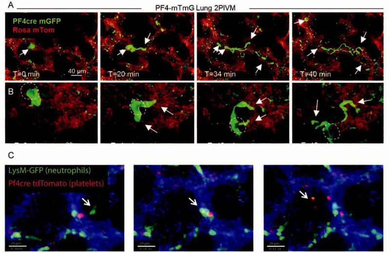
Figure 3. Platelet and Neutrophil Dynamics In The Live Lung.
A and B. Intravital imaging of intrapulmonary megakaryocytes generating proplatelets and releasing them into the lung vasculature. The PF4 promoter (a megakaryocyte and platelet specific marker) was used to drive Cre recombinase expression in membraneTomato membraneGFP (mTmG) reporter mice. When Cre is present in a cell, expression of TdTomato (red) is switched to GFP (green). B. Dark shadow highlights the cell nucleus, establishing its nature as a megakaryocyte as opposed to a conglomeration of platelets. A and B were reprinted with permission from[40]. C. Intravital imaging of mice wherein platelets are labeled in red (PF4-Cre x ROSA-TdTomato) and neutrophils in green (LysM-GFP). Timelapse shows the dynamic formation of a neutrophil-platelet aggregate over the course of 7.5 minutes of imaging. Reprinted with permission from[39].

