
Figure 2.
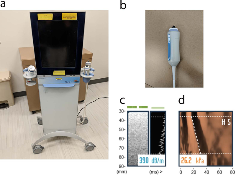
VCTE scanner (a) with the M probe (b) is used to acquire controlled attenuation parameter (CAP, c) and Time Motion (TM) and Amplitude (A) mode shear wave propagation images (d). The CAP estimate of attenuation in units of dB/m is shown in (c). The 5th measurement of liver stiffness in units of Young’s modulus (kPa) is shown in (d). The y-axis is distance from skin, x-axis is time. Slope of the dashed line represents shear wave speed. Median values of these 10 measurements are calculated for stiffness and attenuation assessment. CAP is an integrated technology that quantifies steatosis severity at the same time as liver stiffness assessment on VCTE.