
Figure 4.
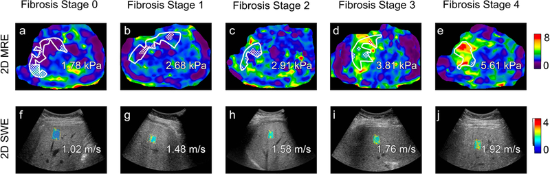
Transverse colorized MR elastograms (3T GE 750 scanner using 2D GRE technique, top) and ultrasound-based two-dimensional shear wave elastography images with placement of regions of interest filled in with colorized elasticity (GE Logiq E9 with C1–6 transducer, bottom) demonstrate increasing stiffness estimates (kilopascals, kPa) or shear wave speed estimates (meters per second) with increasing liver fibrosis stage as determined on histology (Brunt system) in patients with nonalcoholic fatty liver disease. From left to right: stage 0 in 53-year-old man, stage 1 in 49-year-old man, stage 2 in 55-year-old woman, stage 3 in 68-year-old woman, and stage 4 in 72-year-old woman. Regions of interest, an automated 95% confidence grid, and estimated magnitude of complex modulus (“shear stiffness”) values in kPa are overlain on the MR elastograms. Shear wave speed estimates are overlain on the ultrasound images.