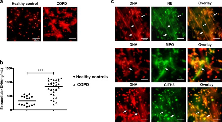
Fig. 2. Excessive NETs in sputum of COPD.
a The immunofluorescence of spontaneous NETs in sputum of COPD and healthy controls. The diluted sputum was seeded on poly-d-lysine-coated coverslips in 24-well round-bottom culture plates and allowed to settle for 1 h without stimulation. Cells on coverslips were fixed with 4% paraformaldehyde and permeabilised with 0.5% Triton X-100. To visualise the backbone of NETs, DNA was stained with propidium iodide (PI) and examined by fluorescence microscopy. Original magnification, ×400. Scale bars = 50 µm. b Concentrations of extracellular DNA in sputum of healthy controls (n = 16) and COPD (n = 32) were detected by PicoGreen fluorescence quantitative assay. Data are expressed as medians. The comparisons were determined by Mann–Whitney test. ***P < 0.001. c The diluted sputum of patient with COPD was seeded on poly-d-lysine-coated coverslips in 24-well round-bottom culture plates and allowed to settle for 1 h without stimulation. Cells on coverslips were fixed with 4% paraformaldehyde and permeabilised with 0.5% Triton X-100. NETs in sputum of COPD were stained with NE, MPO, or CITH3 (green) and extracellular DNA was stained with propidium iodide (PI, red) (NETs: white arrows). Original magnification, ×400. Scale bars = 50 µm

