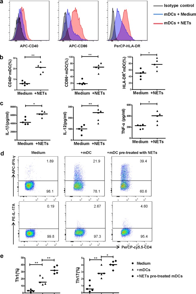
Fig. 5. CSE-induced NETs promote the activation of mDCs and the generation of Th1 and Th17 cells.
a PMNs isolated from patients of COPD were stimulated with 0.3% CSE for 4 h. CSE-induced NETs were harvested by AluI. The purified day 6 monocyte-derived mDCs of healthy donors were stimulated with CSE-induced NETs for 15 h and detected by flow cytometry. Representative histograms of CD40, CD86, and HLA-DR gated from mDCs were shown. b The CD40, CD86 and HLA-DR expression on mDCs with or without CSE-induced NETs stimulation were detected by flow cytometry. Data are expressed as medians (n = 5). The comparisons were determined by Mann–Whitney test. *P < 0.05, **P < 0.01. c The concentrations of IL-1β, IL-12 and TNF-α in supernatants of mDCs with or without CSE-induced NETs stimulation were detected by ELISA. Data are expressed as medians (n = 5). The comparisons were determined by Mann–Whitney test. *P < 0.05. d The purified immature mDCs of day 6 were primed with IFN-γ (10 ng/mL) for 24 h, and stimulated with CSE-induced NETs (20 ng/mL) for another 24 h. CD4+ naïve T cells isolated by negative selection from PBMCs of healthy donors were cultured with mDCs (stimulated with or without NETs) at a DC/T cell ratio of 1:5 for 4 days. Th1 and Th17 in the co-culture condition were detected by flow cytometry. Representative scatter plots of Th1 cells and Th17 cells in the co-culture condition were shown. e The propotions of Th1 cells and Th17 cells in the co-culture condition were detected by flow cytometry. Data are expressed as medians (n = 5). The comparisons were determined by Mann–Whitney test. *P < 0.05, **P < 0.01

