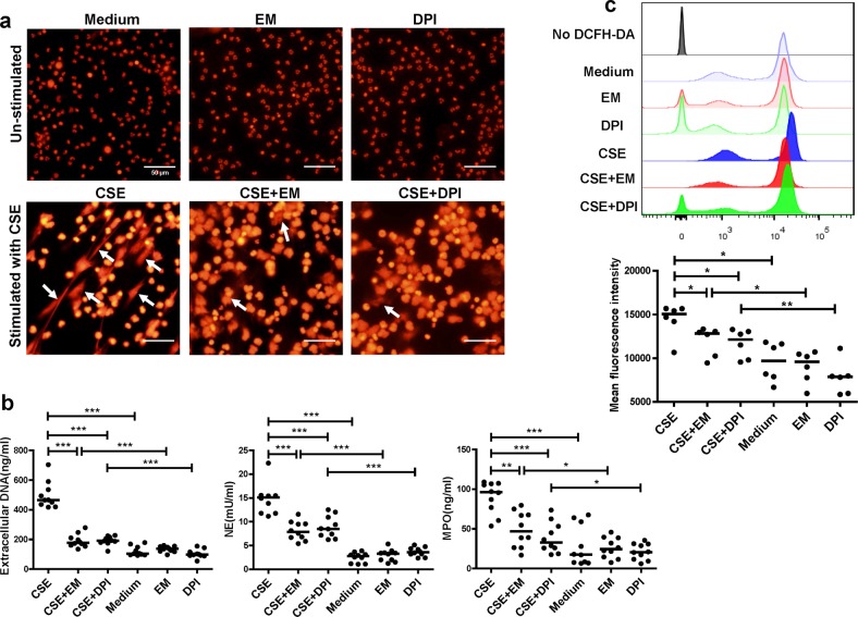
Fig. 6. Erythromycin inhibits CSE-induced NETs drived from human.
a Neutrophils isolated from blood of patients with COPD were stimulated with 0.3% CSE for 4 h at 37 °C ( ± 10 μg/mL erythromycin or ± 5 μg/mL DPI for 30 min). Neutrophils were then fixed with 4% paraformaldehyde for 20 min at room temperature and stained with PI (NETs: white arrows). Original magnification, ×400. Scale bars = 50 µm. b PMNs isolated from patients with COPD were stimulated with 0.3% CSE for 4 h at 37 °C ( ± 10 μg/mL erythromycin or ± 5 μg/mL DPI for 30 min). Quantification of extracellular DNA in supernatants was performed by PicoGreen fluorescence assay. Quantification of NET-associated NE and MPO were detected using the NETosis Assay kit and MPO ELISA kit. Data are expressed as medians (n = 10). The comparisons were determined by Kruskal–Wallis one-way ANOVA on ranks. *P < 0.05, **P < 0.01, ***P < 0.001. c Neutrophils from blood of patients with COPD were pre-treated with erythromycin or DPI for 30 min before CSE stimulation for 1 h. Cells were then stained with 1 μmol/L 2′,7′-dichlorofluorescein diacetate (DCFH-DA) at 37 °C for 30 min and detected by flow cytometry immediately. Intracellular reactive oxygen species (ROS) levels were expressed as mean fluorescence intensity (MFI) of DCFH-DA. Representative histograms of DCFH-DA-dyed PMNs were shown. Quantification of MFI of DCFH-DA were performed by FlowJo v10. The Data are expressed as medians (n = 6). The comparisons were determined by Kruskal–Wallis one-way ANOVA on ranks. *P < 0.05, **P < 0.01

