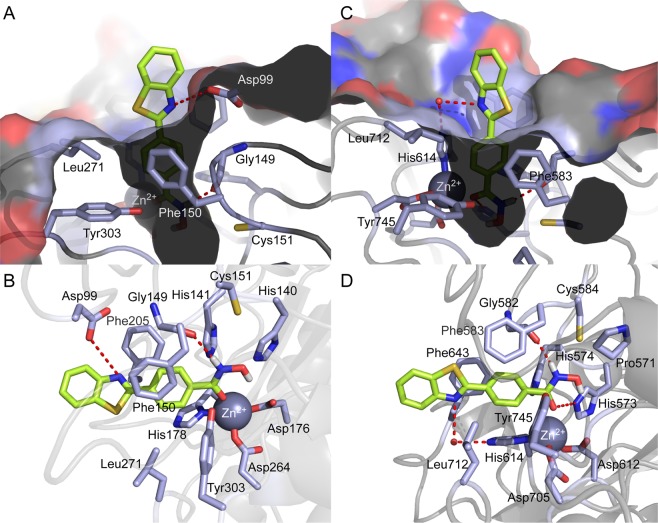
Figure 11.
Docking pose of 9b in the binding site of HDAC1 (PDB code: 4BKX) and HDAC6 (PDB code: 5EF7). (A) Surface representation of HDAC1 and 9b complex. (B) Cartoon and sticks representation of HDAC1 and 9b complex. (C) Surface representation of HDAC6 and 9b complex. (D) Cartoon and sticks representation of HDAC6 and 9b complex. The carbon, oxygen, nitrogen, and sulfur atoms of 9b are shown in lime, red, blue, and yellow, respectively. The side chains of the binding site are colored according to the atom type (carbon, light blue; oxygen, red; nitrogen, blue) and are labeled with their residue name. The hydrogen bond is shown as a dashed line. Molecular docking simulations were performed by Autodock 4.2 and docking poses were visualized using PyMOL1.3.

