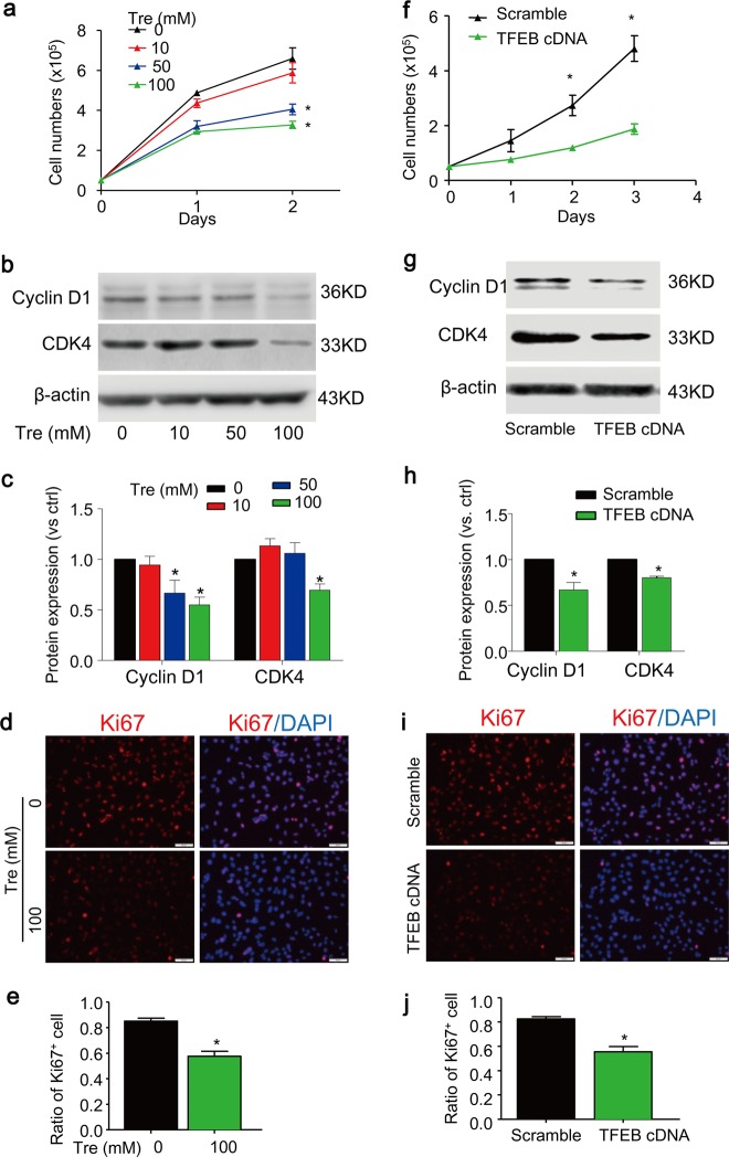
Fig. 2. Trehalose inhibits proliferation of SMCs.
a–e SMCs cultured in full-serum medium were treated with trehalose (0–100 mM) for indicated time points. Cell proliferation was analyzed by counting the cell numbers (a). Immunoblotting analysis (b, c) shows the effects of trehalose (24 h) on the cell cycle protein cyclin D1 and CDK4. d–e Immunofluorescence images show the proliferative cell marker Ki67 (red) in SMCs treated with vehicle or 100 mM trehalose for 24 h. Nuclei were stained with DAPI. f–j SMCs were transfected with scramble or TFEB cDNA plasmids for 24 h and then cultured in full-serum medium for indicated time points. f Cell number counting. g, h Immunoblotting analysis shows the effects of TFEB overexpression on cyclin D1 and CDK4 in SMCs 48 h after transfection. i, j Immunofluorescence images for Ki67 (red) in SMCs transfected SMC SMCs 48 h after transfection. Scale bar = 50 µm *P < 0.05 (n = 4)

