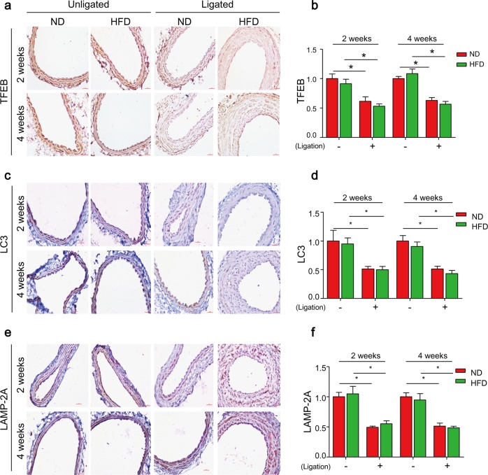
Fig. 5. TFEB-mediated autophagy signaling is defective in arterial media of partial ligated carotid arteries (PLCAs).
The left carotid arteries of mice were partially ligated (ligation) and mice were fed ND or HFD for 2–4 weeks after ligation, the right carotid arteries (unligated) of mice were served as control. a, b Representative IHC analysis of TFEB expression (brown color) in cross sections of carotid arteries. c, d Representative IHC analysis of LC3 expression (brown color) in cross sections of carotid arteries. e, f Representative IHC analysis of LAMP-2A expression (brown color) in cross sections of carotid arteries. Scale bar = 100 μm *P < 0.05 (n = 6–7)

