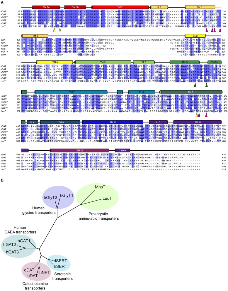
Figure 2.
(A) Multiple sequence alignment of the eukaryotic NSS members with their prokaryotic homolog, LeuT. The colored cylinders represent the transmembrane regions in accordance to the dDAT crystal structure, colored lines represent discontinuous regions within helices and black lines represent the loop regions. Colored triangles highlight the residues involved in the substrate binding subsites A (yellow), B (magenta), and C (green). (B) Phylogenetic analysis of the eukaryotic and prokaryotic NSS members.

