
Figure 1.
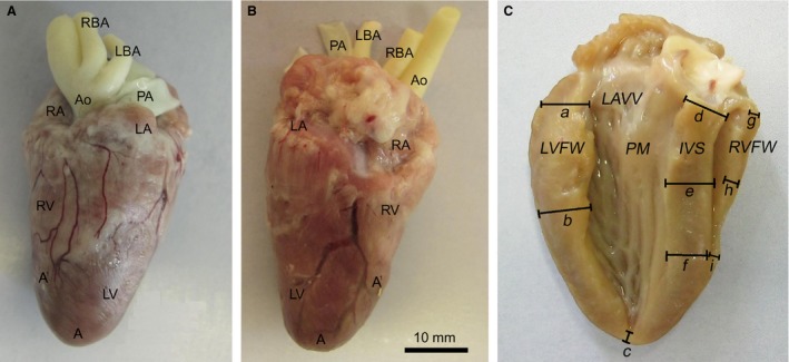
Photographs of an adult chicken heart. The adipose tissue in the coronary sulcus is removed. (A) Ventral surface. (B) Dorsal surface. (C) Illustration of the measuring points for the postmortem evaluation of the myocardial thickness. (a–c) Left ventricular free wall: (a) basal, (b) middle, (c) apical. (d–f) Interventricular septum: (d) basal, (e) middle, (f) apical. (g –i) Right ventricular free wall: (g) basal, (h) middle, (i) apical. A, apex of the heart, A`, apex of the right ventricle; Ao, aorta; IVS, interventricular septum; LA, left atrium; LAVV, left atrioventricular valve; LBA, left brachiocephalic artery; LV, left ventricle; LVFW, left ventricular free wall; PA, pulmonary artery; PM, papillary muscle; RA, right atrium; RBA, right brachiocephalic artery; RV, right ventricle, RVFW, right ventricular free wall.