Figure 1.
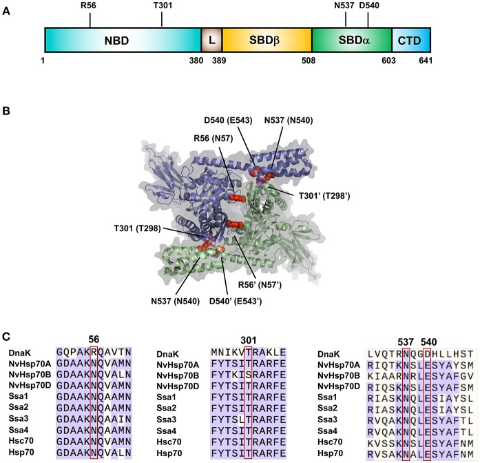
Important residues for Hsp70 dimerization. (A) Location of residues important for DnaK dimerization. (B) Structure of the DnaK dimer based on PDB entry 4JNE (Qi et al., 2013). The two DnaK protomers are colored purple and green. Amino acids important for dimerization of DnaK are labeled with equivalent Hsp70 residues in parentheses in red/magenta. (C) Sequence alignments of major Hsp70 isoforms (bacterial DnaK, Nematostella vectensis Hsp70 (A–C), yeast Ssa1-4 and human isoforms Hsp70 and Hsc70) showing regions critical for dimerization.

