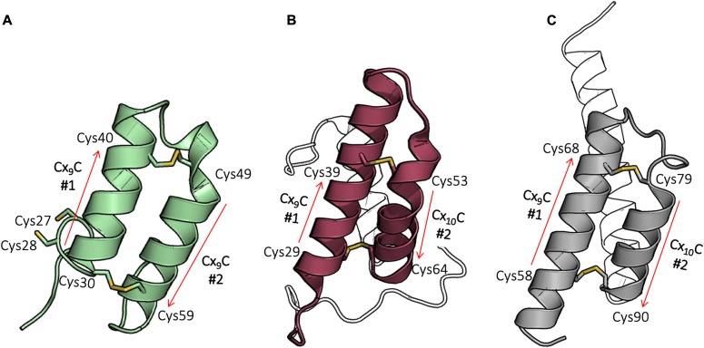
Figure 2. WTCoa6 has a twin CX9C protein fold.
(A) Cox17 (PDB 2RNB): helices are shown in green, and cysteines in the CX9C motifs that form disulfide bonds are shown as yellow sticks. (B) Cox6B (PDB 2EIJ): helices are shown in raspberry, and cysteines in the CX9C (or CX10C) motifs that form disulfide bonds are shown as yellow sticks. (C) WTCoa6 (this work): helices are shown in gray, and cysteines in the CX9C (or CX10C) motifs that form disulfide bonds are shown as yellow sticks. In all panels, the positions of the corresponding motifs are shown as red arrows. In panel (B), part of the N terminus and helix α3 in panels (B) and (C) are not colored for clarity.

