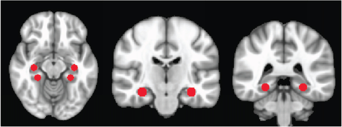
Fig. 1.
Regions of interest delineated on MNI brain. See Table 2 for coordinates. The first image to the left depicts axially the hippocampal (paired, above) and parahippocampal (paired, below) ROIs. The second image depicts the hippocampal ROIs coronally, and the final image depicts the parahippocampal ROIs coronally.

