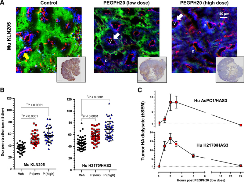
Figure 1.
Enzymatic degradation of tumor HA decreases tumor hypoxia, increases tumor doxorubicin penetration/dispersion, and increases free (nonbound, perfusable) HA within the tumor extracellular milieu. A, Representative images of murine SCC (KLN205) tumors treated i.v. with vehicle or PEGPH20, at 0.0375 mg/kg or 1 mg/kg, highlighting hypoxic regions (visualized with Hypoxyprobe-1, green), blood vessels (anti-CD31, red), and doxorubicin drug penetration (blue); with representative HA-stained (brown) whole tumor sections shown in accompanying insets. Arrows highlight closed (yellow) and open (white) tumor vessels. V, vessels and D, Doxorubicin. B, The area of doxorubicin penetration/dispersion increased in the murine KLN205 with low- (left plot) and high-dose (right plot) PEGPH20. C, Soluble HA fragments appear in the tumor dialysate (tumor interstitial fluid) immediately following PEGPH20 administration (0.0375 mg/kg, i.v.).

