Fig. 3.
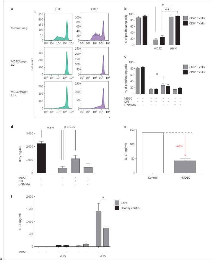
MDSCs from CAPS patients are suppressive on T cells and monocytes. a-c MDSCs from CAPS patients are able to suppress T-cell proliferation in a dose-dependent manner involving ROS. The suppressive effect of MACS-isolated MDSCs (CD66b+) from CAPS patients on T cells (CD4+ and CD8+ T cells) was analyzed by coculture of different MDSC-to-target (PBMC) ratios (1:2 and 1:12) and assessed by CFSE staining and flow cytometry. Shown are representative histograms (a) including a medium control, where PBMCs have been cultured in complete medium only, and the statistics of 3 independent experiments (b); conventional neutrophils (PMNs) did not have any effects on T-cell proliferation. c ROS were blocked by DPI and iNOS by L-NMMA. d, e IFNγ; and IL-17 secretions are decreased by MDSCs. IFNγ; (d) and IL-17 (e) secretions in the supernatant were measured in MDSC/PBMC coculture experiments (see a) by ELISA and multiplex cytokine array. f IL-1β secretion by monocytes is decreased by MDSCs. MDSCs (CD66b+) and monocytes (CD14+) were isolated by MACS from heparinized blood of CAPS patients and cocultured with or without LPS (10 ng/ml) in complete medium at 37°C and 5% CO2. After 4 h of incubation, supernatants were analyzed for IL-1β levels with ELISA. Data are shown as means ± SEM analyzed by a paired t test. * p < 0.05; ** p < 0.01; *** p < 0.001.

