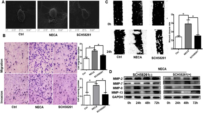
FIGURE 2:
Adenosine promotes GC cell invasion and metastasis through the A2aR pathway in vitro. (A) Representative scanning electron microscopy images showing significantly more pseudofoot and cilia growth. (B) Migration (top) and invasion (bottom) Transwell assays showing increased invasive capability of NECA-treated cells compared with control (Ctrl) or NECA + SCH 58261–treated cells. Scale bar = 100 µm. Bars represent mean ± SEM of at least three independent quadruplicate experiments. (C) Wound-healing assay in MKN45 cells: the scratch was measured 24 h after the treatments. Bars represent mean ± SEM of at least three independent quadruplicate experiments. (D) Immunoblot detection of tumor metastasis–related MMPs. Data are the mean ± SE of triplicate measurements. *, P < 0.05, ANOVA.

