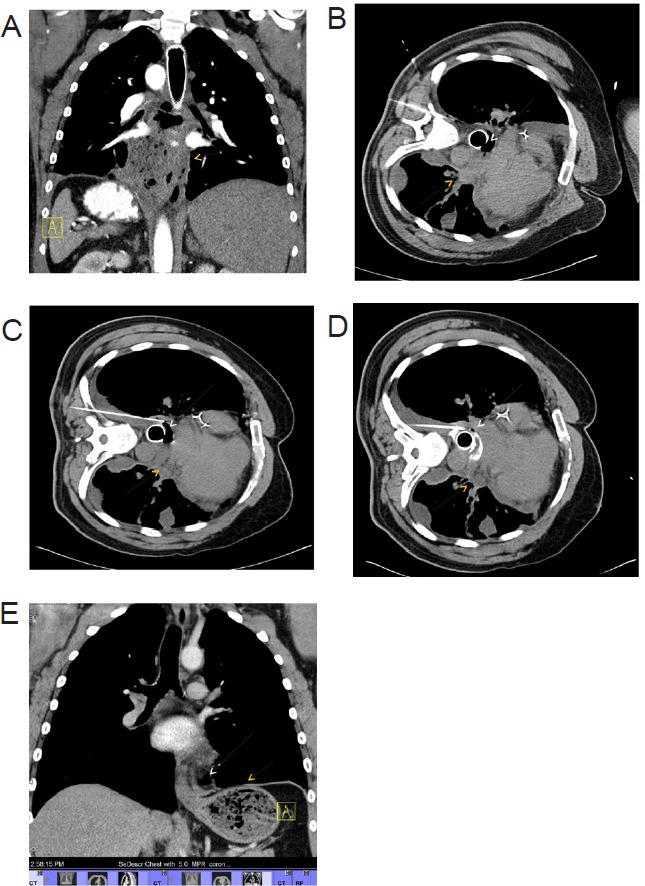
Figure 1.

Computed tomography of chest showing (A) pneumomediastinum with periesophageal abscess extending from carina to GE junction (Yellow arrow), (B) pneumomediastinum (white arrow) with periesophageal abscess (yellow arrow), (C) advancement of pigtail catheter into periesophageal abscess, (D) advancement and dilatation of pigtail catheter into periesophageal and mediastinal abscess, and (E) complete resolution of abscess.
