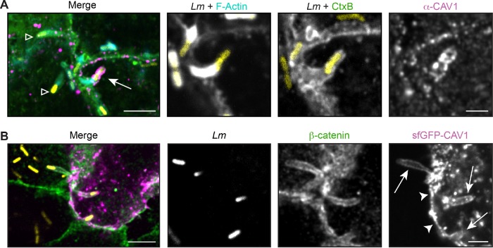
FIGURE 3:
CAV1 localization at the protrusion membrane is not enhanced during L. monocytogenes spread. (A) Infrequent localization of endogenous CAV1 (magenta) in A549 cells. Arrow indicates CAV1-positive protrusion used in inset; open triangle indicates CAV1-negative protrusions. (B) Localization of an sfGFP-CAV1 fusion (magenta) transiently expressed in A549 cells. Similar levels of sfGFP-CAV1 were seen along the plasma membrane (arrowhead) and the protrusion (arrow). For A and B, membranes (green) were detected with CtxB (A) or an antibody to β-catenin (B) after infection with LmTagBFP (Lm; yellow). Scale bar = 5 μm (inset, 2 μm).

