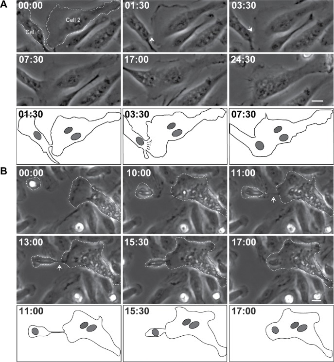
FIGURE 1:
Phase-dense protrusions initiate macrophage fusion. (A) Live imaging of macrophages undergoing type 2 fusion. Macrophages were isolated from the mouse peritoneum 3 d after TG injection and plated on a 35-mm Fluorodish, and fusion was induced by IL-4. Mononuclear macrophage (Cell 1) extends a short phase-dense protrusion (white arrow) toward MGC (Cell 2) immediately before fusion. The bottom panel is a diagram of frames at 1:30, 3:30, and 7:30 min illustrating morphological aspects of the fusion process. In each micrograph, time is shown in minutes:seconds. The scale bar is 10 μm. See also Supplemental Video S1. (B) Macrophage undergoing type 2 fusion extends a long protrusion (white arrow) to initiate fusion. The bottom panels show diagrams of frames at 11:00, 15:30, and 17:00 min. The scale bar is 10 μm. See also Supplemental Video S2.

