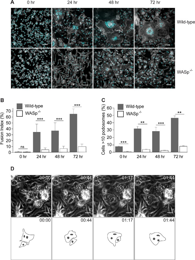
FIGURE 6:
WASp is required for macrophage fusion in vitro. (A) Fusion of WT and WASp-deficient macrophages at various time points after the addition of IL-4. After 24, 48, and 72 h, cells were fixed and labeled with Alexa Fluor 488–conjugated phalloidin (white) and DAPI (teal). The scale bars are 50 μm. (B) The time-dependent fusion indices for WT and WASp-deficient macrophages. Results shown are mean ± SD from three independent experiments. Three to five random 20× fields per sample were used to count nuclei (100–150 nuclei/field; total 1500 nuclei). ***p < 0.001. (C) The time-dependent podosome formation in fusing macrophages. The fraction of cells with >10 podosomes for each time point was calculated. Four random 20× fields each containing ∼200–300 cells were used to count podosomes. **p < 0.01, ***p < 0.001. (D) Live imaging of IL-4–treated WASp-deficient macrophages. In each micrograph, time is shown in hours:minutes. A rare fusion event detected in the population consisting of ∼1200 macrophages is shown.

