FIGURE 7:
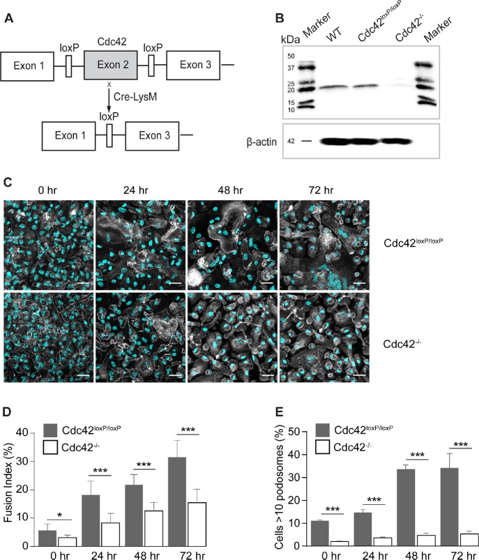
Loss of Cdc42 in macrophages results in impaired fusion in vitro. (A) Schematic diagram of the generation of a myeloid cell-specific Cdc42-/- mouse. Conditional gene-targeted mice with exon 2 of Cdc42 gene flanked by a pair of loxP sequences (Yang et al., 2007) were crossbred with LysMcre mice to allow Cdc42 gene excision in myeloid cells. (B) Cdc42 deletion in isolated macrophages was examined by SDS–PAGE (11% gel) followed by Western blotting using anti-Cdc42 rabbit monoclonal antibody (ab187643; Abcam). (C) Fusion of WT and Cdc42-deficient macrophages at various time points after the addition of IL-4. Cells were fixed after 24, 48, and 72 h and labeled with Alexa Fluor 488–conjugated phalloidin (white) and DAPI (teal). The scale bars are 50 μm. (D) Time-dependent fusion indices of WT and Cdc42-deficient macrophages. Results shown are mean ± SD from three independent experiments. Four to five random 20× fields per sample were used to count nuclei (300 nuclei/field; total ∼4050 nuclei). *p < 0.05, ***p < 0.001. (E) The time-dependent podosome formation in fusing macrophages. The fraction of cells with >10 podosomes for each time point was calculated. Four random 20× fields each containing ∼200–300 cells were used to count podosomes. ***p < 0.001.

