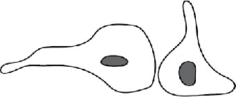
TABLE 2:
Patterns of macrophage fusion.
| Pattern mediated by short protrusionsa | Schematic showing cell polarity | Type 1 fusion | Type 2 fusion |
|---|---|---|---|
| Leading edge to the cell body |  |
34% | 31% |
| Leading edge to rear edge |  |
23% | 18% |
| Leading edge to leading edge |  |
20% | 45% |
| Other | 23% | 6% |
aSee also Supplemental Videos S1–S5.
