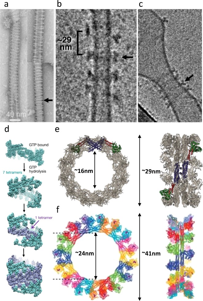
FIGURE 2:
Dynamin helices and rings. (a) Left: A continuous dynamin-1 helix arranged on lipid nanotubes, in the presence of GTP-γS (Marks et al., 2001). Reproduced with permission from Nature Publishing Group. Right: same as left after incubation with 500 µM GTP for 30 min. Note the increase in the helical pitch (arrows) and possible discontinuous helices/rings. (b) DRP1 helical polymers incubated with GTP, leading to an increase in helical pitch (Mears et al., 2011). Reproduced with permission from Nature Publishing Group. These structures (arrow) could represent helices with a very elongated pitch, or rings of DRP1. (c) Rings of MxA observed on a lipid template, as seen in Kochs et al. (2005); Haller et al. (2010). Reproduced with permission from Academic Press and ASBMB. (d) A model for dynamin-1–based membrane fission, as described by Kong et al. (2018). Reproduced with permission from Nature Publishing Group. (e) Models for rings of DRP1 (Kalia et al., 2018) and (f) of MxA (Haller et al., 2010). Reproduced with permission from ASBMB.

