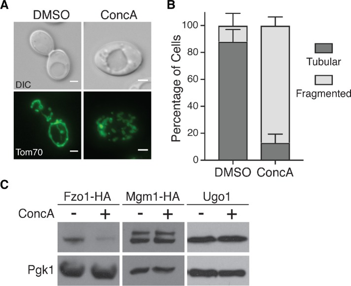
FIGURE 2:
Vacuole impairment induces mitochondrial fragmentation and Fzo1 depletion. (A) Representative maximum-intensity projection images of wild-type cells expressing Tom70-GFP treated with DMSO or concanamycin A (ConcA) for 6 h. Scale bar = 2 μm. (B) Quantification of mitochondrial phenotypes (tubular or fragmented) from panel A. Error bars show mean ± SE of three replicates. n = 100 cells/replicate. (C) Whole cell extracts from yeast cells expressing the indicated protein grown in the absence or presence of concanamycin A (ConcA) for 4 h were analyzed by Western blot with anti-HA, anti-Ugo1, and anti-Pgk1 antibodies.

