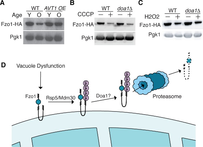
FIGURE 7:
Vacuole-dysfunction–induced Fzo1 degradation occurs in response to elevated metabolic stress. (A) Whole cell extracts from young (Y) and old (O) wild-type or AVT1 overexpressing Fzo1-HA cells were analyzed by Western blot with anti-HA and anti-Pgk1 antibodies. Age ranges (n = 50 cells): WT, Y = 0–2, O = 7–9; AVT1 OE, Y = 0–2, O = 7–10. (B, C) Whole cell extracts from wild-type (WT) and doa1Δ yeast expressing Fzo1-HA grown in the absence or presence of carbonyl cyanide m-chlorophenyl hydrazone (CCCP) (B) or hydrogen peroxide (H2O2) (C) for 4 h were analyzed by Western blot with anti-HA and anti-Pgk1 antibodies. (D) Illustration showing that Fzo1 is degraded by a proteolytic cascade requiring Doa1 and redundant actions of SCFMdm30 and Rsp5 in response to vacuole impairment.

