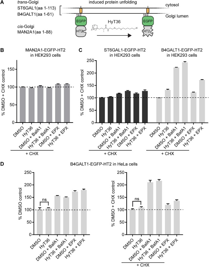
FIGURE 1:
Degradation profiles of Golgi-localized model QC substrates. (A) Domain outline of Golgi QC substrates targeted to the cis- (MAN2A1-EGFP-HT2) and trans- (ST6GAL1- and B4GT-EGFP-HT2) Golgi. (B) Flow cytometry analysis quantifying the levels of MAN2A1-EGFP-HT2 in HEK293 cells on the indicated treatments for 6 h (EPX, epoxomicin; BafA1, bafilomycin A). CHX was added 1 h prior to the indicated treatments. (C) Same analysis as in B for HEK293 cells expressing B4GT-EGFP-HT2 or ST6GAL1-EGFP-HT2 (n = 2, data represent mean ± SEM). (D) Flow cytometry analysis quantifying the levels of B4GT-EGFP-HT2 in HeLa cells on the indicated treatments for 6 h in the absence (left) or presence (right) of CHX (n = 3, data represent mean ± SEM, results of t test are shown).

